Whole Body Ultrasound
What is an ultrasound?
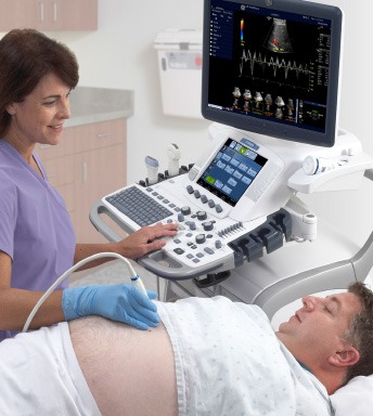
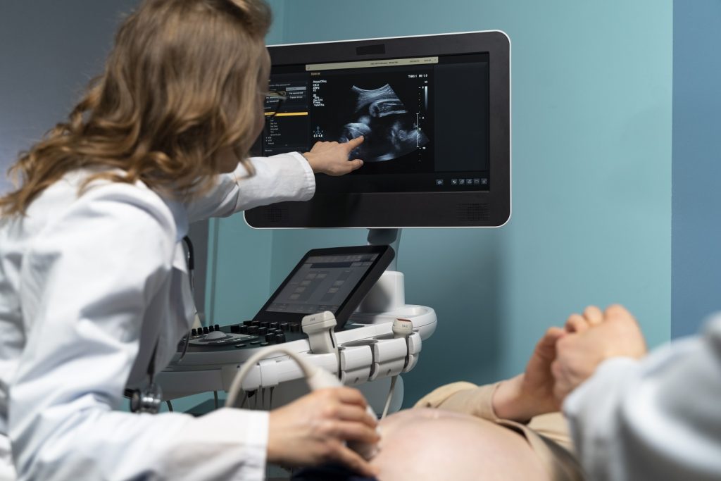
Ultrasound is the term used for high-frequency soundwaves. Ultrasound examinations use these sound waves to produce a picture or image onto a screen showing the inside of your body. An ultrasound is carried out by a Radiologist using a smooth, hand-held device called a transducer that they move across the body with a sliding and rotating action. The transducer transmits the high-frequency sound waves into your body. Different sound waves are reflected from different soft tissue, structures or parts in the body in different ways. These sound waves are converted to electrical impulses that produce a moving image displayed on a screen.
An ultrasound has many advantages. It is painless and does not involve radiation, which means it is very safe. The high-frequency sound waves ensure images show very high detail, capable of looking at the very tiniest parts of the body. Ultrasound can be carried out while there is movement, so it is excellent for the imaging of babies and children.

Whole body ultrasound:
There are many reasons an ultrasound may be requested. For example, it is used to examine organs in the abdomen and other parts of the body. Colour Doppler ultrasound can be used to watch blood flow in any of the arteries or veins throughout the various parts of your body. High-resolution ultrasound can be used to evaluate the musculoskeletal system (muscles, bone- and joint-related). Breast ultrasound is an important part of the assessment of any breast lump. Ultrasound can take high-quality images of most parts of your body, which makes it an excellent diagnostic test.
How do I prepare for an ultrasound?
This will depend on the type of ultrasound that is requested. Listed below are some of the common examinations, with the preparation generally required.
All ultrasound examinations:
Wear clothing that will provide easy access to the area that is being imaged.
Bring any previous radiology examinations you have had with you (including ultrasound, magnetic resonance imaging, X-ray and computed tomography scans), so that they can be used for comparison and assessment.
Specific ultrasound examinations:
Abdomen ultrasound: You will usually need to fast (have nothing to eat or drink) for 4-6 hours before the examination. This ensures there is no food or fluid covering the area that is to be examined. It also ensures the gall bladder is expanded to provide a clearer image.
Breast ultrasound: No preparation is required.
Female pelvis ultrasound

Pelvic / Gynaecology Ultrasound Scan
What is a pelvic or gynaecological ultrasound?
This is an ultrasound that examines the organs of the female pelvis. This includes the ovaries, the cervix, the uterus/womb and the lining of the uterus (referred to as the endometrium).Why might I need a pelvic ultrasound?
Common reasons for this are:- Pelvic pain or painful periods
- Heavy periods or abnormal bleeding
- Pelvic mass
- Infertility
- Postmenopausal bleeding
- Pain or bleeding following delivery of a baby or after a miscarriage
- Assessment of IUCD/Mirena position
What can be seen on the scan?
- Abnormalities with the uterus such as fibroids
- Ovarian cysts, masses or polycystic ovaries
- Abnormalities with the endometrium like polyps or abnormal thickening
- Abnormal fluid adjacent to the uterus or ovaries
How is it performed?
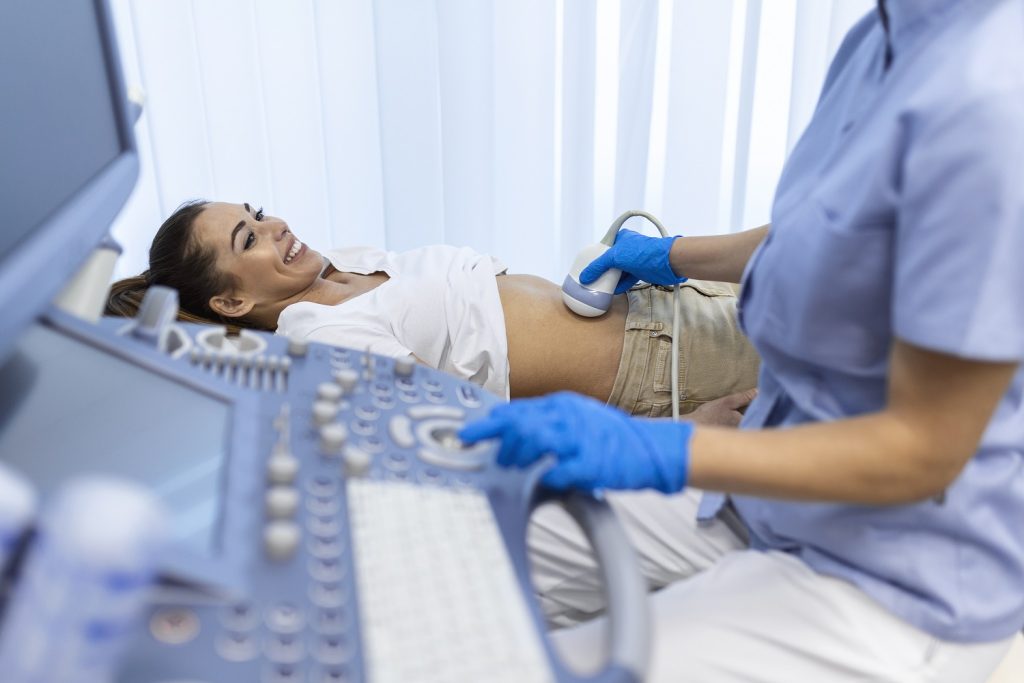
Both transabdominal and transvaginal scanning is recommended for adequate views of your pelvic structures. Transabdominal scanning is performed by placing a probe and gel on the skin of your lower abdomen. This provides a good overall assessment of pelvic organs and not a lot of detail. A transvaginal scan involves a sterilized, covered probe being gently placed into the vagina whilst you are covered. This usually provides clearer images of the pelvic structures as this probe lies closer to the uterus, ovaries, and cervix. It is less uncomfortable than a PAP smear. You will always have the choice about whether this is performed.

Do I need a full bladder?
Fluid in the bladder allows clearer images to be obtained during transabdominal scanning. It allows a “window” for the scan and moves your bowel out of the pelvis helping better visualisation. Your bladder needs to be partially filled (your bladder should not be so full that it causes you pain). Emptying the bladder two hours before the examination and drinking ¾ of a litre of water before your appointment is advised. You will be allowed to empty your bladder before the transvaginal scan begins.
Thyroid ultrasound: No preparation is required.
Testes ultrasound: No preparation is required.
Musculoskeletal (muscle-, bone- and joint-related) ultrasound: No preparation is required.
Renal (kidney-related) ultrasound: You will need to drink 750 mL of water 1 hour before the ultrasound. Do not go to the toilet after drinking the fluid. Drinking the water will enlarge the bladder, enabling it and the surrounding internal areas to be examined.
Renal (kidney) arteries ultrasound: You will need to fast (have nothing to eat or drink) for 8 hours before the examination to ensure that the renal arteries are not covered by food or fluid.
What are the risks of an ultrasound?
Ultrasound is a safe examination that provides excellent imaging without any significant risk to the patient.
What are the benefits of an ultrasound?
Ultrasound provides excellent imaging of the soft tissues of the body and is a safe procedure that does not have the risks associated with imaging that uses radiation. There are no proven harmful effects of sound waves at the levels used in ultrasound when carried out in a proper clinical setting, such as private radiology practice or hospital.
Ultrasound can be used with patient movement, so is ideal for imaging babies and children. Imaging movement is also very valuable in musculoskeletal (muscles, bone- and joint-related), gynaecological (women’s health, especially of the reproductive organs) and vascular (blood vessel-related) ultrasound. Dynamic imaging (moving pictures) provided by images using ultrasound sound waves gives the opportunity to look at the inside of the body in positions or with movements where there is pain or movement restriction.
Rarely, a specific ultrasound contrast medium is injected into a vein of the arm to detect certain types of diseases or problems. If the radiologist feels that this will be useful, then this will be explained to you at the time of the examination.
Ultrasound is mostly non-invasive, provides accurate imaging tests of the body, is readily available and is relatively inexpensive.

