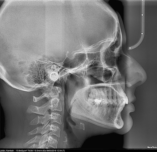
Lateral Cephalogram
Another useful assessment tool in dental imaging is the lateral cephalogram. This is an x-ray that generates a side view of the head that can provide important information about the teeth and jaw. The lateral cephalogram shows the facial structure, bone, and soft tissue. Your radiologist or dental practitioner can use these images to study the relation of your teeth to your jaw, assess problems in alignment or growth patterns, and prepare treatment.
The lateral cephalogram is best for observing jaw alignment. If you have an overbite or underbite, for example, these images will provide better detail and allow your practitioner to develop the best approach for your needs. This x-ray is commonly used in children to predict growth patterns in the teeth and jaws. The lateral cephalogram is used with adults as well, to study changes made from treatment or track other issues.
If you are working with an orthodontist, the lateral cephalogram may be ordered alongside the OPG. This x-ray is useful in tracking the progress of treatment.
What to Expect With a Lateral Cephalogram
An appointment for a lateral ceph should take approximately 15-20 minutes. Often, you’ll receive this scan in conjunction with the OPG. You will need to remove jewellery and metallics from your head and neck. For your lateral ceph you’ll be asked to keep your teeth together, and may lean your forehead against a steadying implement. The x-ray itself will be completed in only a few seconds

