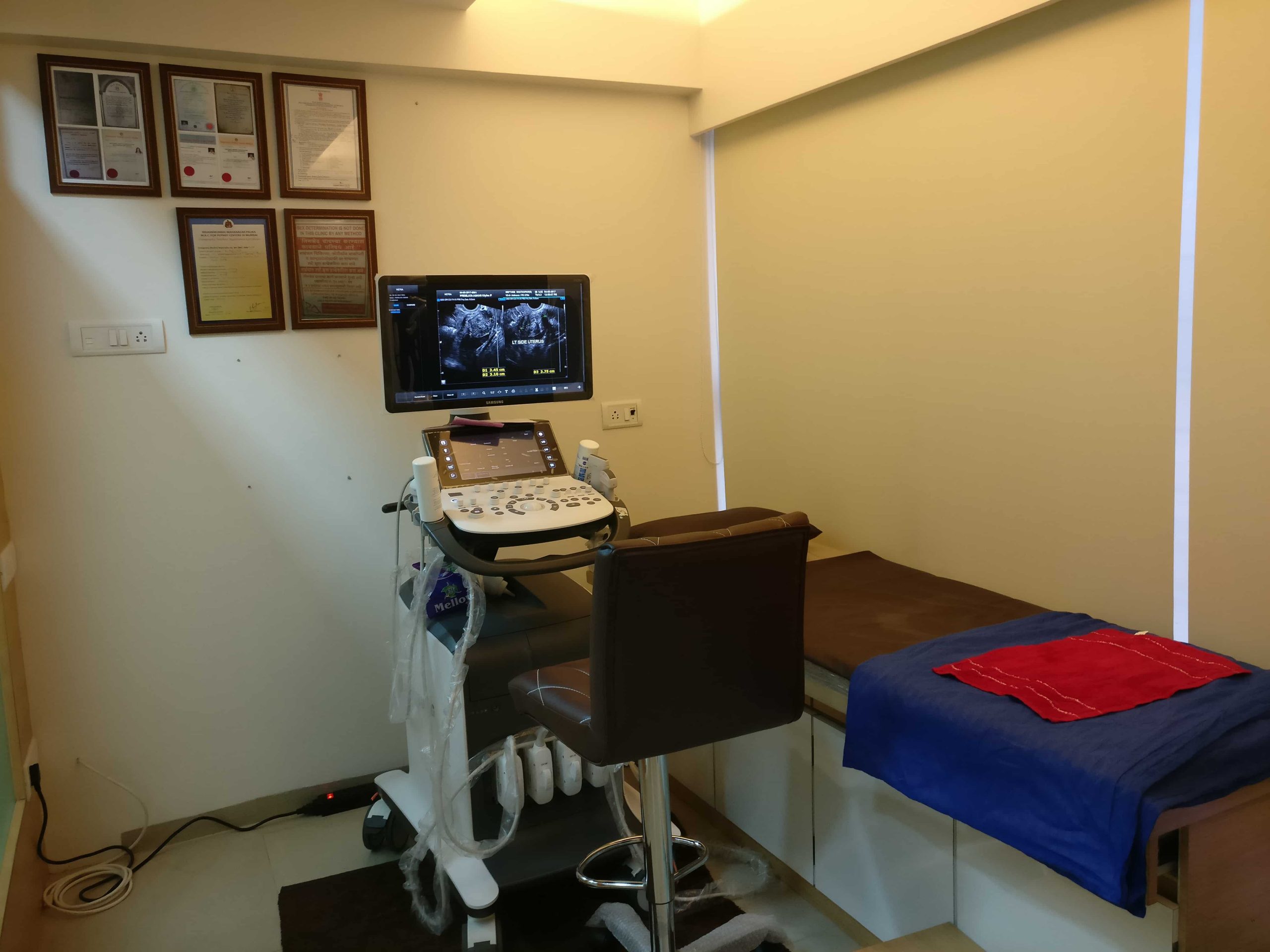
Colour Doppler Examinations
Upper Limb Arterial Duplex Ultrasound
May be one or both sides, according to the clinical history and specific clinicians request.
Indications
Evaluation of known or suspected peripheral vascular disease, aneurysm or pseudoaneurysm, subclavian steal syndrome or uneven brachial pressures
Patient Preparation
None required
Patient history
Previous vascular surgery, history of diabetes, smoking, CVA/TIA, previous imaging
Aorto-iliac Duplex Ultrasound
This examination may be performed on its own, or in conjunction with a unilateral or bilateral lower limb duplex examination, depending on the specific clinical history.
Indications
Abdominal bruit, evaluation of aneurysm or pseudoaneurysm, evaluation of inflow of peripheral vascular disease, follow-up of surgical or interventional procedures
Patient Preparation
Fast for 6 hours prior to examination.
Patient history
Previous vascular surgery, history of diabetes, smoking, CVA/TIA
Lower Limb Arterial Duplex color Doppler
This examination may be unilateral or bilateral, and will always include examination of the aorta and iliac arteries (See previous section – Aorto-iliac Duplex Ultrasound).
Indications
Claudication, rest pain, ulceration, ischemic changes, evaluation of aneurysm or pseudoaneurysm, follow-up of surgical or interventional procedures.


Patient Preparation
None required
Patient history
Symptoms, previous vascular surgery, history of diabetes, smoking, CVA/TIA, previous imaging
Lower limb Venous Doppler
Indications:
Ultrasound can be used to assess for the presence of Deep Vein Thrombosis (DVT), and Chronic Venous Insufficiency (CVI) or varicose veins. Symptoms may include swelling, pain, and redness in the legs.
Examination:
Ultrasound gel is applied to the skin on your legs and an ultrasound probe is moved over the skin surface.
During the DVT examination, the technologist may need to repeatedly press firmly on your skin, and squeeze your calf or foot. This is considered an urgent examination.
The CVI study is undertaken in a similar manner to the DVT examination, however, it generally takes a little longer to perform. This is not considered an urgent examination.
Preparation:
None required
Penile Duplex Doppler Ultrasound
Utility of the ultrasound:Penile Doppler Ultrasound testing is an extremely useful investigation in erectile dysfunction (ED) and Peyronie’s Disease. It is a sophisticated test to objectively measure the blood flow in and out of your penis. This test will give important information as to the nature of penile structure and function, both in the erect and non-erect state.
It can, therefore, help to identify the underlying cause of ED – i.e. the Doppler US can determine if there is an arterial problem (blood inflow), a venous leak problem (blood outflow) or both. Arterial disease in the penis diagnosed on this ultrasound may be related to a more diffuse arterial disease such as in the heart.
If you have Peyronie’s Disease it will also assess the nature of the plaque and scar tissue in the penis. This will help to determine if you are a candidate for certain therapies or operations. In addition, it will allow assessing your penile curvature directly.
How is the test done?It involves the use of specialized ultrasound to see inside the penis and to look at the internal structures, the arteries, and the veins. In order to get the maximum information, you will need some medication to give you an erection. This also simulates “real-life”. This medication is injected by a tiny needle into the penis. The medication then works automatically over about 10 minutes. Depending on the condition of your penis, you may need more than 1 injection. This is the only part of the test which may be a little uncomfortable. Most men describe the sensation as “like a hair being pulled out the back of my hand”. The whole process is over in about 3 seconds.
Once Doctor has determined you have a satisfactory response from the injection, he will then assess for any deformities of the penis. A doctor will then perform the ultrasound and will identify and measure the arterial inflow, the venous outflow, and the penile integrity.
What are the risks of the test?
The biggest potential risk of this study is an erection which lasts for too long (called a priapism). This condition can cause damage to the penis. We have a strict protocol which means that you will only go home once we confirmed that the erection has subsided.
How long will the ultrasound take?
You will be with at clinic for about 1 hour. This takes into account the injection and the ultrasound.


Carotid Doppler
Indications:
To assess the Carotid Arteries for plaque (“thickening” of the arteries), and blockages. Reasons for investigation include a history of stroke, smoking, high cholesterol, and where there is known vascular disease elsewhere in the body.
Preparation:
None required.
