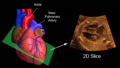
Fetal Echocardiography
Fetal echocardiography consists of the assessment of baby’s heart before birth. Most children have a normal heart. Therefore, most heart scans prior to birth show no obvious abnormalities and the major heart structures can be identified as expected in a normal fetal heart.
Approximately 8 in 1,000 babies born alive will show a heart defect. Half of these are major defects that can be identified before babies are born if a specialised scan of the fetal heart is performed. Although most complex abnormalities are well tolerated by the baby while in utero, treatment is usually required after birth and maybe as early as in the first few days of life, often due to changes in the way the blood goes round following delivery.
Minor abnormalities such as small holes in the heart may also be detected, but they do not disturb the way the heart works, even after birth. Treatment is usually unnecessary for minor abnormalities, but follow up may be needed as appropriate. Apart from structural defects, some babies will need to be assessed for rhythm disturbances as well.
Fetal echocardiography is usually carried out between 22-24 weeks of gestation and a single scan is generally adequate to assess the fetal heart. However, in some situations, the fetal heart can be assessed as early as 13-14 weeks of gestation and provide a lot of information and reassurance to some parents who are at risk of having a child with a congenital heart defect. In these cases, a second fetal echo is scheduled later on in pregnancy.
You do not need to have a full bladder and you do not need to drink before the examination.

